Down Syndrome Baby Ultrasound Picture
Excess skin in the back of the neck nuchal translucency A shorter-than-normal femur thigh bone. A missing nose bone.
Diagnosis Of Down Syndrome Youtube
And d position negative in a trisomy18 fetus at 23 5 weeks.

Down syndrome baby ultrasound picture. The most predictive finding of Down syndrome on prenatal ultrasound is an absent nasal bone. See down syndrome baby stock video clips. If you put the two tests together the detection rate can be as high as 85 per cent.
So adding in these additional markers will take the average detection rate of the standard technique of 80 up to 95. There are multiple prenatal genetic screening strategies and diagnostic tests aimed at accurate prenatal identification of Down Syndrome and other aneuploidies. What is the chance of my baby will have down syndrome.
Browse 488 down syndrome baby stock photos and images available or search for down syndrome kids or down syndrome newborn to find more great stock photos and pictures. 1011 down syndrome baby stock photos are available royalty-free. 8 years experience Obstetrics and Gynecology.
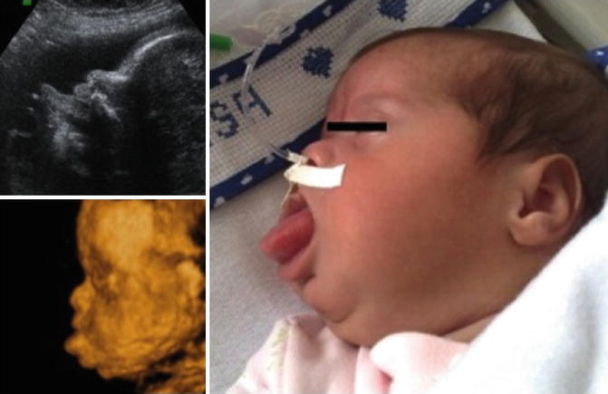
Baby down down baby down syndrome newborn down syndrome love down syndrome down syndrome handicapped child isolated child with down syndrome child syndrome down syndrome child. On an ultrasound an image of a developing fetus also called a sonogram visible signs a baby may have Down syndrome include. However they are seen more frequently in fetuses with an abnormality.
Soft markers are sonographic findings that do not in themselves cause any adverse outcomes. Little baby with Down syndrome hid under blanket and smiles slyly Portrait of a down syndrome baby girl smiling with happiness and play with camera. 2690 down syndrome baby stock photos vectors and illustrations are available royalty-free.
Down Syndrome trisomy 21 is the most common chromosomal disorder in live born infants. Approximately 30 of babies with Down syndrome have detectable abnormalities on the mid-trimester ultrasound 1. About 6000 babies are born with Down Syndrome each year in the United States or about 1 in every 700 births.
Ultrasonography is the imaging modality mainstay of prenatal screening and diagnosis of Down syndrome and it is often used in combination with biochemical tests. Babies with Downs syndrome are more likely to have a small or absent nose bone with a flat profile. C position positive in a fetus with Down syndrome at 28 2 weeks.
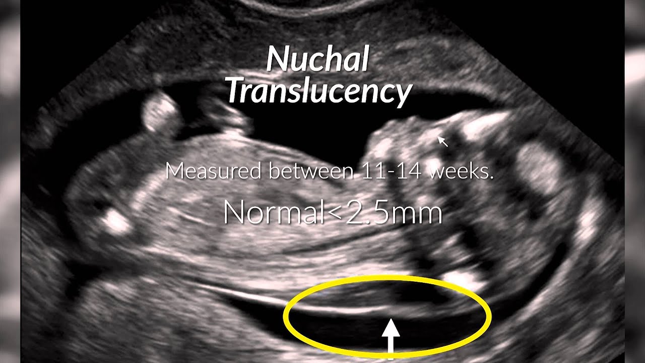
They often have leakage across the tricuspid valve and reverse flow in the ductus venosus. They explained that our childs nuchal fold or the fluid at the back of the babys neck was developing far too thick a possible indicator of Down Syndrome. Ultrasound pictures of baby with down syndrome A 34-year-old female asked.
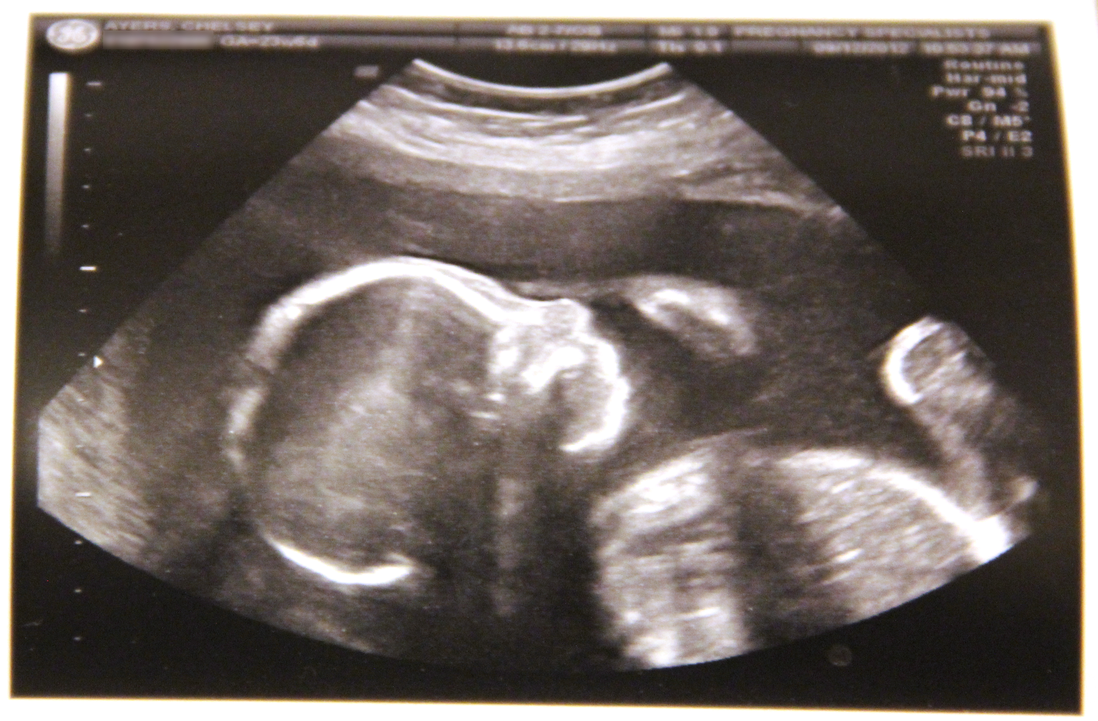
An ultrasound also know as a sonogram is a test done during pregnancy which uses sound waves to generate a picture or image of the fetus. Shortened humerus in a fetus may be associated with high risk for Down syndrome. A position zero in a euploid fetus at 24 6 weeks gestation.
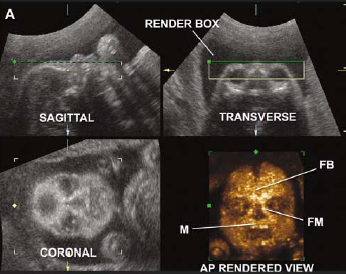
Occasionally but not always infants with Down syndrome show subtle signs on an ultrasound that can make your doctor suspect that the fetus has Down syndrome. Twodimensional ultrasound images of fetal profile FP line at. Ultrasound image reproduced with permission from Measuring the Arm on the World Wide Web.
Thickened nuchal fold nuchal translucency Duodenal Atresia double bubble Echogenic bowel Cardiac heart anomalies Choroid plexus cyst Echogenic intracardiac focus. It does not mean you are likely to have a Down syndrome baby if the scan is outside the normal range. The Nuchal cord translucency ultrasound alone probably picks up about 75 per cent of babies with Down syndrome and the blood test alone about 60 per cent.
Down syndrome son playing at home - down syndrome baby stock pictures royalty-free photos images. See more ideas about ultrasound sonography diagnostic medical sonography. It also means that some Down syndrome babies are not detected by this scan and method of testing.
Jan 22 2021 - Explore Drmobeen siddiquis board Down syndrome ultrasound on Pinterest. This screen is shown to be able to identify the majority of Down syndrome babies. The following are ultrasound markers that are seen more frequently in fetuses with Down syndrome.
My trisomy is 114. B position zero in a fetus with Down syndrome at 21 3 weeks. This does not mean your baby will have Down syndrome however.
/babyboyultrasound-7bf2ced4b4794754b67dea974b7ec744.jpg)
What To Look For In Your Baby Boy Ultrasound

Do Babies With Down S Syndrome Have No Nose Bone Babycentre Uk

Ultrasound Of Baby At 10 Weeks With Down Syndrome Page 1 Line 17qq Com

How Doctors Test For Down Syndrome Parent

Ultrasound Images Of A Normal Fetus A And A Fetus With Trisomy 21 B Download Scientific Diagram
Pin On Obstetrică Patologica

Soft Markers Down Syndrome April 2018 Birth Club Babycenter Canada
Down Syndrome The Hudson James

Finding Down Syndrome Via Ultrasound Little Doctors

Down Syndrome Screening Chromosomal Abnormality Screening Ultrasound Care

Pin On Prenatal Down Syndrome Diagnosis

Fetal Facial Profile Markers Of Down Syndrome In The Second And Third Trimesters Of Pregnancy Vos 2015 Ultrasound In Obstetrics Amp Gynecology Wiley Online Library
Prenatal Diagnosis Of Beckwith Wiedemann Syndrome Using 3d Ultrasound And Fetal Mri Springerlink

Baby Nuchal Translucency Scan Center In Chennai Nuchal Translucency Down Syndrome Ultrasound Prenatal Screening

Diagnosis Of Fetal Syndromes By Three And Four Dimensional Ultrasound Is There Any Improvement

Down Syndrome Ultrasound Page 1 Line 17qq Com
Prenatal Ultrasounds To Help Identify Potentially Devastating Illnesses And Conditions Baby Glimpse

Genetic Sonography 3d Us

Ultrasound Scan Spots Down S Syndrome Nature News
Post a Comment for "Down Syndrome Baby Ultrasound Picture"